What is the medial patellofemoral ligament (MPFL)?
The medial patellofemoral ligament (MPFL) plays a crucial role in the intricate web of soft tissues that stabilize the knee. It serves as the link between the inner portion of the kneecap (patella) and the elongated thigh bone (femur), collectively forming the patellofemoral joint.
Injuries to the MPFL typically transpire when the patella experiences dislocation or partial dislocation (subluxation), often due to traumatic incidents during sports or accidents. Such injuries can also result from naturally lax ligaments, which are more commonly observed in females, or due to variations in an individual's bone structure. Individuals with these injuries are often described as having patellar instability.
What is MPFL reconstruction?
MPFL reconstruction is a surgical procedure designed to establish a fresh medial patellofemoral ligament, effectively enhancing knee stability and safeguarding the joint against further harm. This surgical solution is particularly well-suited for individuals who have encountered recurrent instances of dislocation.
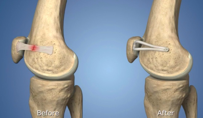
Injury to the MPFL
In a healthy knee, the components of the patellofemoral joint move harmoniously as the knee flexes and extends. The patella smoothly glides within the femur's trochlear groove. The medial patellofemoral ligament (MPFL) is instrumental in maintaining the patella's alignment within this groove, acting as a leash that restrains excessive movement.
When a patellar dislocation occurs, it can cause damage to the surrounding soft tissues as the patella momentarily "jumps" out of its track and then forcefully returns to its place. Typically, dislocations lead to a tear in the ligament on the inside of the knee, the MPFL.
If left untreated, an injured MPFL may heal naturally but often in a lengthened and weakened state. This can result in ongoing instability, making the patella more susceptible to future dislocations. Recurrent dislocations can lead to cartilage damage within the knee, which poses a more complex challenge. Damaged cartilage increases the risk of developing patellofemoral arthritis, a condition that is considerably more challenging to manage. Therefore, seeking treatment to prevent further patellar dislocations is highly advisable.
What is the reconstruction protocol for MPFL?
Patients who experience patellar instability undergo a comprehensive assessment that encompasses a physical examination and a review of their medical history. To gain a more comprehensive understanding of the condition of the cartilage in the patellofemoral joint and determine the most suitable treatment approach, a magnetic resonance imaging (MRI) scan is typically performed. The MRI helps in evaluating whether the patient is a candidate for MPFL reconstruction or requires a bony procedure like tibial tubercle transfer.
While non-operative treatments have a limited role in addressing patellar instability, patients who have encountered a single dislocation without any cartilage damage on MRI may undergo short-term immobilization and physical therapy. However, the majority of candidates for MPFL reconstruction have experienced multiple dislocations. (MPFL reconstruction may be considered in cases of a single dislocation only if there are additional knee problems necessitating surgical intervention.)
In MPFL reconstruction, the damaged ligament is replaced with a portion of the patient's own tendon. The procedure is performed arthroscopically, allowing the surgeon to visualize the surgical area through small incisions. Typically lasting about an hour, the surgery is performed as an outpatient procedure, and patients return home on the same day with their knee stabilized in a brace.
MPFL reconstruction yields excellent results with a very low rate of complications. Although rare, potential complications may include fractures, infections, or blood clots. Notably, this procedure can be safely performed in children with open growth plates (the areas where bones continue to grow), making it suitable for young patients who require treatment.
What to expect after MPFL reconstruction?
Following MPFL reconstruction, patients are typically allowed to bear weight on the affected leg immediately after the procedure. A brace is provided, which must be worn for approximately six weeks. This brace maintains the leg in a straight position during walking. To prevent the development of scar tissue and joint stiffness during the ligament's healing process, patients use a continuous passive motion machine (CPM) at home. The CPM machine facilitates movement of the patellofemoral joint without requiring muscle engagement. Once the quadriceps, the major muscle in the thigh, has regained sufficient strength to support the joint, patients can commence physical therapy, typically around six weeks post-surgery.
In most cases, individuals can anticipate a return to sports or physical activities within a range of 4 to 7 months following MPFL reconstruction. However, it's essential to note that recovery times can vary, influenced by factors such as an individual's anatomy, healing capacity, and overall health prior to the surgery.