Osteochondral Autograft Transfers
Our joints are equipped with a resilient, smooth substance known as cartilage. This essential surface facilitates the effortless and painless gliding of joints, granting us the freedom to move and flex. However, as the natural aging process unfolds or as we engage in sports and physical activities, this cartilage can begin to deteriorate and sustain damage. This deterioration can give rise to specific conditions that manifest as knee joint pain, stiffness, and swelling.
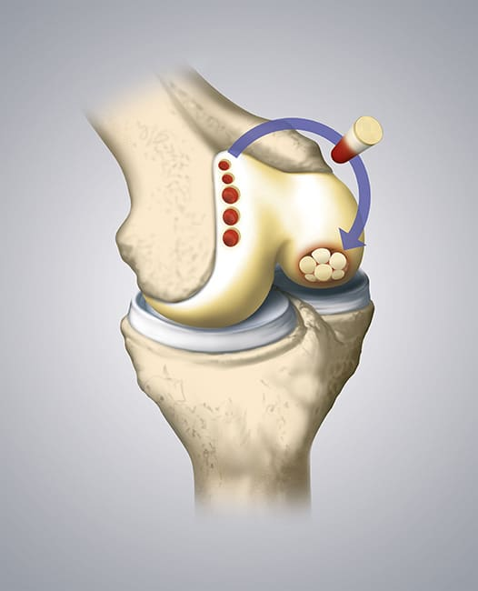
One of the notable procedures designed to alleviate these issues is osteochondral autograft transfer (OATS). In this surgical technique, a small plug consisting of both bone and cartilage is extracted from a less weight-bearing area of the knee using a specialized cylindrical coring device. This plug is then transplanted into an area of the knee that exhibits a symptomatic osteochondral defect. Typically, OATS procedures are performed arthroscopically. It's worth noting that the target defect areas are usually around 1 cm or smaller to minimize any potential complications at the donor site where the plug was removed.
While microfracture procedures are often recommended for addressing minor defects, there are instances where microfracture may not be the most suitable option. This includes scenarios involving patients who are taking blood thinners, individuals with cysts beneath the cartilage defect area, or those engaged in physically demanding occupations or sports that require a faster post-surgery return to activity.
In most cases, a small open incision is made at the location where the plug is being implanted. This ensures precise placement that aligns with the normal contour of the remaining articular cartilage. While the majority of the surgery can be conducted arthroscopically, it does necessitate several arthroscopic incisions around the knee to facilitate the transplantation of the donor graft into the recipient site.
Post-Operative Protocol for Osteochondral Autograft Transfers
Patients who undergo osteochondral autograft transfers typically follow a specific rehabilitation protocol to optimize their recovery. Initially, they are instructed to refrain from bearing weight on the affected leg for approximately 6 weeks following the surgery. During this period, a continuous passive motion (CPM) machine becomes an essential component of their rehabilitation routine, and patients are encouraged to use it for 6-8 hours each day. The CPM machine aids in moving the patellofemoral joint without relying on the patient's muscles, which is especially important during the initial stages of healing.
Once the 6-week mark post-surgery is reached, patients are granted permission to gradually increase their weight-bearing capacity, as tolerated. This phase also marks the commencement of using a stationary bike as part of their rehabilitation program, contributing to the restoration of their knee's functionality. Overall, the goal is to ensure that patients regain their strength, experience no swelling or pain during physical activities, and see a return to their normal level of function. This comprehensive process typically spans a minimum of 5-6 months to achieve a full recovery. It's essential to understand that recovery times can vary depending on individual factors, such as anatomy, healing capacity, and overall health prior to the surgery.
Take the first step towards better health - Book your appointment now